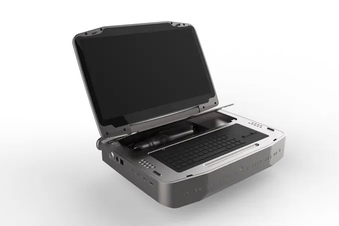
Fundus imaging is a cornerstone of modern eye disease diagnosis. With advances in technology, ultra-wide-angle and handheld fundus camera systems, such as the Portable Ultra-Wide-Angle Fundus Camera (PUWF), are redefining how clinicians capture high-quality retinal images efficiently—whether in hospitals, mobile clinics, or community screening programs.
Ultra-wide-angle fundus imaging captures a vast view of the retina—often over 130° field of view (FOV)—allowing clinicians to visualize both the central and peripheral retina.
Detects peripheral retinal lesions that traditional cameras may miss
Enables early diagnosis of retinal diseases
Supports comprehensive retinal assessment in a single capture
Ultra-wide-angle imaging is especially valuable in pediatric and neonatal ophthalmology, where full retinal visualization is critical for early intervention.
Modern handheld fundus cameras provide both portability and advanced imaging capabilities. Key benefits include:
Broader retinal coverage for complete retinal screening
Enhanced peripheral pathology detection for early intervention
Improved workflow in neonatal and pediatric imaging
Portability for bedside exams, mobile clinics, and community outreach programs
The PUWF model, for example, uses medical-grade sensors and LED illumination to deliver true-color, high-resolution images, even in non-dilated patients, ensuring diagnostic accuracy in diverse clinical settings.
Handheld and portable fundus cameras allow eye care providers to:
Perform exams in NICUs, mobile clinics, and outreach programs
Integrate with telemedicine platforms for remote diagnosis
Share images instantly with specialists for faster referral and treatment
Reduce barriers to quality retinal care in underserved or rural areas
Portable fundus imaging ensures that diagnostic care reaches patients wherever they are, improving both accessibility and outcomes.
Handheld ultra-wide-angle fundus cameras are particularly effective in:
| Clinical Application | Benefit |
|---|---|
| Retinopathy of Prematurity (ROP) Screening | Full retinal view for early intervention in neonates |
| Diabetic Retinopathy Monitoring | Detect microaneurysms and hemorrhages across the retina |
| Age-Related Macular Degeneration (AMD) Tracking | Monitor macular changes over time |
| Glaucoma Progression Assessment | Visualize optic disc changes and peripheral damage |
By combining advanced imaging with portability, clinicians can increase screening coverage while maintaining high diagnostic confidence.
With advances in imaging sensors, ultra-wide-angle optics, and AI integration, handheld fundus cameras are becoming essential tools for:
Ophthalmology departments
General medical practices
Neonatal and pediatric clinics
Mobile and telemedicine screening programs
The handheld fundus camera is no longer just a convenience—it is a critical instrument for modern retinal care, improving accessibility, workflow, and patient outcomes.


